Optipedia • SPIE Press books opened for your reference.
Photoreceptors
Excerpt from Field Guide to Visual and Ophthalmic Optics
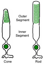
 Two types of photoreceptors reside in the retina: cones and rods. The cones are responsible for daytime vision, while the rods respond under dark conditions. The cones come in three varieties: L, M, and S types (for long, middle, and short wavelength). Each cone type responds to a different portion of the visible spectrum, allowing for color vision. Rods have a spectral sensitivity that differs from the cones. Photoreceptors are specialized cells for detecting light. They are composed of the outer nuclear layer that contains the cell nuclei, the inner segment that houses the cell machinery, and the outer segment that contains photosensitive pigment. The outer segment of a rod has discrete disks saturated with rhodopsin molecules, while the outer segment of a cone contains similar photosensitive molecules in a series of folds. The outer segment absorbs photons, which initiates an electrochemical transmission through the cells and retinal nerve fibers, up into the brain.
| Cones | Rods |
| Color Vision | Monochromatic |
| No sensitivity in the dark | High sensitivity in the dark |
| Respond in bright light | Bleached in bright light |
| Slow temporal response | Fast temporal response |
| Mostly in fovea | Mostly in periphery |
| Some in peripheral retina | None in fovea |
| High visual acuity | Low visual acuity |
| In fovea, one neuron per cone | Many rods per single neuron |
Cone diameter is roughly 2.5 μm in the fovea and rapidly increases outside fovea to 10 μm in periphery. Rod diameter is roughly 3 μm at a field angle of 18° and increases in size to 5.5 μm in periphery. The central 200 μm of the retina is free of rods. The total number of cones in the retina is 6.4 million. There are roughly 125 million rods in the retina.
J. Schwiegerling, Field Guide to Visual and Ophthalmic Optics, SPIE Press, Bellingham, WA (2004).
View SPIE terms of use.

