Imaging the Cancer Cure
New medical imaging technologies may accelerate the early detection of cancers.

The imaging community has an important role to play in advancing the way cancer is studied, diagnosed, and treated. This work includes understanding how cancer cells move and migrate, picking up cancers at an earlier stage than current diagnostics can, or predicting and monitoring a patient's response to treatment.
To drive this forward, the US National Cancer Institute in 2016 launched its Cancer Moonshot campaign, supported by then President Barack Obama, to accelerate cancer research. The National Photonics Initiative (NPI) responded to this with a white paper and cancer technology road map that identifies the most promising existing and new technologies for increased and concerted investment.
"I think that it's being recognized that the quality of imaging is going to play a major role in reducing cancer mortality over the next decade," says Thomas M. Baer, a past chair of the NPI steering committee and executive director of the Stanford Photonics Research Center (USA). "That was part of the message that we tried to send to the White House.
"There's clear evidence that to develop imaging technology that allows early detection in certain types of cancer, such as lung cancer, what's needed is funding for clinical trials to develop the technology." The NPI, a collaborative alliance among industry, academia, and government seeking to raise awareness of optics and photonics, has also committed to leverage more than $3 billion in annual private investments for cancer research from over 350 hospitals, patient-advocacy groups, and the scientific and medical technology communities for early detection technologies for the most aggressive cancers.
The funding comprises two components, Baer says. "One is to fund the clinical trials and deployment of technology that already exists. The other is to look at new applications and new technologies for different types of cancer that remain to be effectively addressed in terms of early detection or any intervention.
"However, I don't want to build up the expectations that there's a breakthrough in technology that's going to significantly change the mortality rate of cancers because this doesn't do anybody any good," Baer says. "There's an evolution in technology that looks like it is going to have a reasonable impact, and it is important to be deployed."

Technologies impact patients across the cancer treatment spectrum, from prevention and early detection to treatment. (NPI image)
Lung scans could save lives
Baer is adamant that investment in photonics is extremely important and could save lives. "But we're not on the verge of an immediate breakthrough in any one specific area of cancer treatment or early detection," he adds. "The one exception would be lung cancer, where I think there is now preliminary evidence that spiral CT scans of lung cancer in at-risk people will have a major impact on mortality."
A spiral CT scan uses a computer linked to an x-ray machine to make a series of detailed pictures of areas inside the body.
The x-ray machine scans the body in a spiral path. This allows more images to be made in a shorter time than with older CT methods. A dye may be injected into a vein or swallowed to help the organs or tissues show up more clearly on the x-ray.
Spiral CT scans also create more detailed images and may be better at finding small, abnormal areas inside the body.
Advances with quantitative imaging
Computed tomography (CT) based imaging is the dominant tool for both lung cancer detection and to evaluate the status of drug or radiotherapy response. But that is being replaced by quantitative imaging, the process of using medical imaging as a measurement tool rather than just providing pictures. Quantitative imaging is moving into routine clinical application as well as lung cancer screening.
The National Comprehensive Cancer Network (NCCN) and other groups have developed nodule-size thresholds to guide routine lung cancer screening practice. "Image-acquisition processes are continuously improving resolution," James L. Mulshine, vice president for research at Rush University Medical Center (USA), explains. "Software systems are becoming more facile relative to efficient workflow integration and standardized reporting formats.
"In the future, I would expect the routine performance of quantitative imaging to be done with high-quality image acquisition so that fewer images would be adequate for robust quantitation. This would allow clinicians to have a higher confidence that they are making clinical decisions based on accurate data," Mulshine says.

Melanoma cells in red, demarked by a fluorescent protein. The green denotes functional lymphatic vessels, which are the conduits that spread melanoma throughout the body. This is in a mouse model of melanoma. Because the lymphatic system houses the local immune system, this type of technology is essential for research to improve upon checkpoint blockade immunotherapies, a focus of the Moonshot. (Image: S. Kwon, UTHealth)
Fast hyperspectral imaging
While quantitative imaging is showing promise in lung cancer detection, it is another technology, hyperspectral imaging, that is being developed for cancer of the gastrointestinal (GI) tract. A team at University of Cambridge (UK), led by SPIE member Sarah Bohndiek, has developed a hyperspectral-imaging-enabled endoscopy system to detect and characterize early cancerous changes in the GI tract.
"Hyperspectral imaging is a technique that allows you to record an image that includes not only spatial information, but also spectral information, relating to the color of light coming back from the object that you're imaging," Bohndiek explains.
"The reason that we're interested in this is because at the moment, there's a lot of limitations with endoscopies, which are just done with white light."
The technique essentially replicates what your eye could see, "but our eyes aren't perfect," she says. "They only sample red, green and blue, in terms of their color spectrum. They're not looking at very detailed spectral analysis, and because of that, the contrast for picking up very early cancer in the GI tract is low and leads to very high miss rates for early diagnosis."
To date, application of the technique in endoscopy has been very limited because of the instrumentation challenges. "Normally when carrying out spectroscopy, you do a point source of illumination, and you interrogate a single point and sample," Bohndiek says. "You collect the spectral information on your camera, and then you move that point around and build up a picture. If you're doing that in endoscopy, it is going to mean the patient's got to lie there for three hours. It's a very low-throughput technique."
Details about a new bimodal multispectral endoscope developed by Bohndiek and her colleagues at Cambridge were presented in a paper, "A multispectral endoscope based on spectrally resolved detector arrays," at the OSA/SPIE European Conferences on Biomedical Optics earlier this year. The scope allows for reflectance imaging in the visible spectral region and fluorescence imaging in the near infrared (NIR).
SPIE Senior Member Eva Sevick-Muraca, chair of the US Cancer Moonshot task force of the US National Photonics Initiative, presented a poster on the role of photonics enabled imaging in cancer research, prevention, and treatment at a conference in June, marking the first anniversary of the US Cancer Moonshot initiative. Sevick- Muraca's technical poster was titled, "National Photonics Initiative: Cancer Moonshot Task Force: Medical Imaging Used to Accelerate Progress."
Measuring tumors in color
Another challenge is with data analysis, being able to process data fast enough that it could display information back to the endoscopist in near real time.
Over the last few years, the traditional color camera has evolved from having a two-by-two array of color filters to a three-by-three or four-by- four array of color filters. So you can sample more than just three colors; you sample 16 or 25 colors. By building up that information, you can distinguish the different molecules in the body that have different absorption properties and different colors.
The interesting thing about that is you can measure the oxygenation of tissues, and tumors have different levels of oxygenation, typically, than normal tissue. You can also measure things such as the metabolic rate in the tissue, and tumors tend to have an altered metabolism compared to normal tissue.
Multi-parametric MRI
In the detection and treatment of prostate cancer, there are two major problems. First, the screening test, Prostate Specific Antigen (PSA), has many false positives, leading to unnecessary biopsies. Second, the subsequent biopsies have a high false negative rate.
The lack of accuracy of current diagnostic methods can lead to overtreatment, with its associated risk of impotence or incontinence. It can also lead to missing cancers that should have been treated.
Studies show that multi-parametric MRI (mpMRI) improves individual risk assessment to better identify who needs treatment and who does not. At the same time, it reduces the number of biopsies, along with costs and patient risk of infection. Studies also show that use of targeted Positron Emission Tomography (PET) tracers can detect small metastases that typically go undetected, thereby making a major change in treatment decisions and likelihood of efficacy.
"Multi-parametric MRI is more than just an anatomical picture of the nodules; it can characterize the tumor tissue for likelihood of cancer," says, Richard Frank, chief medical officer of Siemens Healthineers North America. "This mpMRI pattern can direct the biopsy needle to targets within the heterogeneous tissue with the greatest risk of being cancer, to the nodules - and the regions within the nodules - so that the most relevant tissue samples are obtained."
For mpMRI, the technology is in place and a standardized reading and reporting system (PI-RADS) is in use already. What remains is to make it more widely available.
The technology for PET is also in place. The main challenge is completion of the multiple clinical trials necessary for regulatory approval.
To permit the development of algorithms that support the human reader and therapeutic decision-maker, it is necessary to capture data from the variety of non-interoperable electronic medical records and create a cloud-based infrastructure, or Data Lake. "This will enable artificial intelligence, including "Deep Learning" and natural language processing, to capture, store, integrate/assimilate, analyze, and share all of the data coming in," Frank adds.
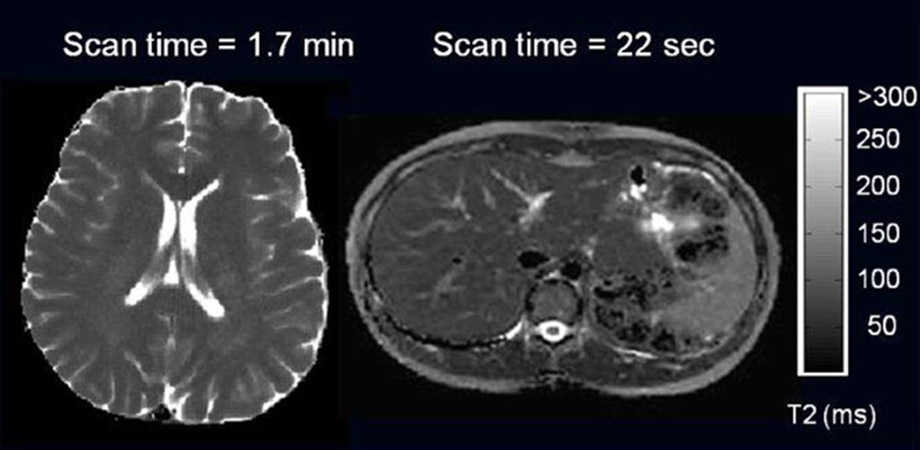
Spin-spin relaxation time (T2) maps of (left) brain, (middle) liver, and (right, color map) the left-ventricular heart wall obtained from highly undersampled MRI data acquired with a radial fast spin-echo sequence and reconstructed using a model-based algorithm. The combined radial acquisition and reconstruction strategies allow acquisition of data with high spatiotemporal resolution within a short scan time. (SPIE Newsroom)
Miniaturized falloposcopes
When it comes to detecting ovarian cancer, the current screening tools are manual examination, pelvic ultrasound, and blood tests to detect the CA-125 protein, the biomarker that is found in greater concentration in tumor cells than in other cells of the body.
"To detect cancer early, you need to use the sensitivity and resolution of optics and optical imaging techniques to be able to detect cancer when it's not invasive, when it's at a very early, curable state," says SPIE Fellow and SPIE Board Member Jennifer K. Barton of the University of Arizona Cancer Center (USA). "It can't be done with an MRI or a CT scan."
The types of tumors that can be detected on whole-body techniques are quite large, and usually the cancer is quite advanced by the time you can detect it there. "Optics is highly sensitive, has great resolution, but its challenge is that it can't penetrate through the whole body," says Barton, who received the 2016 SPIE President's Award. "Light can't penetrate, so we need to get our sensor and our light sources right up next to the tissue that we want to look at."
Barton's tiny, highly flexible endoscope, or falloposcope, is a wandlike imaging device that combines several optical imaging techniques to detect ovarian cancer in the fallopian tubes, where many researchers believe the cancer originates. Her team has already tested rigid prototypes in pilot clinical studies, but the new flexible version will not require any incisions, making it more suitable for screening.
"Ideally, we go through orifices or tubes that we already have in the body, and one of the most challenging ones that you can think of is the fallopian tubes," Barton says. "We can make a sub-millimeterdiameter endoscope that can travel through the uterus into the fallopian tubes and reach the ovary without having to make any incisions in the tissue.
This is enabled by recent advances in fiber-optics and portable light sources as well as, higher-power and more capable lasers. The idea of building light-based endoscopes isn't new, but because of all the advances in technology, we've really been able to push the limits on sensitivity and the capability and do extreme miniaturization."
Raman spectroscopy
The ability to dispense with biopsies and diagnose cancer on the spot would be a huge boost for doctors. Researchers at Leibniz Institute of Photonic Technology (IPHT) in Jena (Germany) have been doing just that with the development of a handheld fiber-optic probe that can be used to perform multiple non-linear imaging techniques without the need for tissue staining.
By adopting photonic approaches, researchers can monitor live processes in cells or tissue on a molecular level. Also, light allows the diagnosis and treatment of diseases in a gentle way and opens a pathway to minimally invasive medicine.
In this regard, spectroscopic methods such as fluorescence, IR absorption, and Raman are particularly noteworthy. They have the potential to provide a clinician with adequate support in the form of clinically relevant information under both ex-vivo and in-vivo conditions. Spectroscopic methods offer the decisive advantage of obtaining molecular information directly from the examined cells or tissue.
Using spectroscopic methods, it is possible to obtain qualitative and quantitative biochemical information in addition to morphological information that can be correlated with clinical results.
Typical examples of basal cell carcinoma (BCC) by raster scanning Raman micro-spectroscopy (RMS). Scale bar: 400 microns. H&E: Hematoxylin and eosin staining. Inflamed D: Inflamed dermis. (SPIE Newsroom)
"One particularly efficient method in this regard is Raman spectroscopy allowing one to monitor molecular vibrations distinct for every molecule," says SPIE Fellow Jurgen Popp, director at IPHT. "A vibrational spectrum can, therefore, be interpreted as a type of characteristic molecular fingerprint of examined cells or tissues. Spectral contributions are assigned to proteins, lipids, nucleic acids, and carbohydrates."
As cancer and other pathologic anomalies are accompanied by changes in the biochemical composition and structure of biomolecules, the Raman spectrum provides a sensitive and specific fingerprint of the type and state of the specimen.
Advantages of the technique for biomedical problems are that it is both label-free and non-destructive. The combination of spectral and lateral information creates a powerful imaging technique.
"Our own work predominantly focuses on developing and using Raman approaches for characterizing biological cells and tissue with the aim of providing sensitive and selective tools to potentially complement established clinical diagnostic methods," Popp adds. "Currently we have achieved a stage of development beyond mere basic feasibility."
However, these approaches are far from being integrated into routine clinical diagnostics. "Optical spectroscopic approaches have proven their potential as regards to certain diagnostic issues in numerous proof-of-principle tests," Popp says. Their actual effectiveness now needs to be tested under routine clinical conditions in patients or in comparative studies using samples, he says.
-Mark Venables is a freelance technical editor and writer.
This article was originally published in the October 2017 edition of SPIE Professional magazine.
Related SPIE content:
Photonics steps up for Cancer Moonshot effort
A decade's worth of advances in five years is the goal of the photonics community and partners, working on imaging, diagnostics and data management.
Jennifer Barton: OCT and fluorescence spectroscopy lead to early cancer detection tools
The BIO5 Institute at the University of Arizona promotes collaborative, interdisciplinary work in biosciences.
Extending the capabilities of quantitative MRI
A technique based on highly undersampled radial MRI yields data with excellent spatiotemporal resolution for quantitative parameter mapping without increasing scan time.
Multimodal spectral imaging speeds cancer diagnosis during surgery
A biophotonics technique based on autofluorescence imaging and Raman scattering can diagnose skin tumors during surgery faster than conventional histopathology, without tissue sectioning or staining.
Fast Raman imaging of living cells for biomedical applications
A profiled laser allows for dynamic Raman imaging of living cell structure and functions, and alkyne tags improve the Raman signal with minimal physiological perturbation.
Single lens system for forward-viewing navigation and scanning side-viewing optical coherence tomography
The optical design for a dual modality endoscope based on piezo scanning fiber technology is presented including a novel technique to combine forward-viewing navigation and side viewing OCT.


