New laparoscopic imaging technique accurately maps biological tissue for minimally invasive surgery
Laparoscopy, a minimally invasive surgical technique, has become the standard of care for many procedures such as prostatectomies and appendectomies due to its benefits, including faster recovery times, reduced scarring, and lower medical costs. However, the technique faces challenges in visualization, particularly when it comes to identifying critical anatomical structures, assessing tissue perfusion, and distinguishing cancerous tissue. The limited field of view (FOV) and poor contrast in laparoscopic images often lead to subjective decision-making based on the surgeon’s experience, which can result in wide variability in clinical outcomes. These challenges have spurred research into optical technologies that provide more accurate, objective guidance for surgeons.
As reported in the Journal of Biomedical Optics (JBO), a team of researchers from Johns Hopkins University has developed a new laparoscopic imaging tool that addresses these challenges by using advanced techniques to map tissue properties with high precision. The device integrates stereo depth estimation and speckle-illumination spatial frequency domain imaging (si-SFDI) to produce detailed optical property maps of tissues. Unlike traditional methods, this system offers a quantitative approach to imaging by measuring key properties such as absorption and scattering, which can differentiate healthy from cancerous tissue with high sensitivity and specificity.
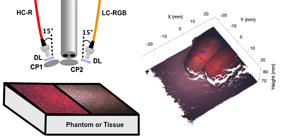
The new system uses a compact two-camera laparoscope equipped with a fiber-coupled laser to generate high-contrast speckle patterns on the tissue surface. These patterns allow the system to estimate optical properties without the need for numerous images. The system results in improved measurement accuracy, particularly for complex tissue geometries. In contrast to earlier versions of si-SFDI, which required 10 or more images, the new technique can obtain accurate data with just two image frames, enabling future real-time mapping during surgical procedures.

Durr Computational Biophotonics Lab at Johns Hopkins University has demonstrated the potential to map the 3D profile and quantitative optical properties of complex surgical scene-like geometries. Graduate researcher Anthony Song is shown operating the optical setup. Photo courtesy of A. A. Song (Johns Hopkins University).
“Using a compact multimode fiber to generate laser speckle patterns makes speckle illumination spatial frequency domain imaging a compelling method for measuring wide-field quantitative optical properties over a large field of view, even in physically constrained settings such as minimally invasive surgery,” remarks Song.
This new method provides a simple yet powerful tool for surgeons to obtain quantitative optical property maps during laparoscopic procedures, potentially reducing reliance on subjective assessments and improving clinical outcomes. By offering detailed tissue information, it has the potential to enhance the precision of minimally invasive surgeries and help identify critical tumor margins.
For details, see the original Gold Open Access article by A. A. Song et al., “Speckle-illumination spatial frequency domain imaging with a stereo laparoscope for profile-corrected optical property mapping,” J. Biomed. Opt. 30(S1), S13710 (2025), doi: 10.1117/1.JBO.30.S1.S13710
| Enjoy this article? Get similar news in your inbox |
|