New imaging technique to improve head and neck cancer surgery

Head and neck squamous cell carcinoma (HNSCC) ranks as one of the most common cancers globally, with over 650,000 new cases reported each year. Surgical intervention is often the primary treatment, but surgeons face a difficult challenge: they must completely remove the cancer while preserving as much surrounding healthy tissue as possible. This balance is crucial, as damage to nearby nerves can lead to significant post-surgical complications, affecting patients' quality of life.
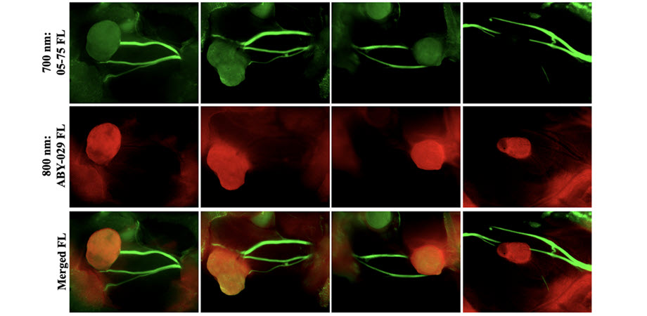
A team of researchers from Oregon Health & Science University and Thayer School of Engineering at Dartmouth has made strides in addressing this challenge by developing a new imaging technique that enhances the visibility of both tumors and nerves during surgery. As reported in the Journal of Biomedical Optics (JBO), their study focused on using fluorescence-guided surgery (FGS) with two different near-infrared (NIR) fluorophores—one specific to tumors and another to facial nerves.
In experiments using a human HNSCC xenograft model, the researchers successfully demonstrated that the two types of fluorophores could be used together to clearly differentiate between cancerous tissues and nerves. The nerve-specific fluorophore showed no significant interference from surrounding tumor tissues, ensuring accurate nerve visualization even in the presence of cancer.
This advancement in FGS offers a promising tool for surgeons, potentially improving their ability to perform cancer resections while minimizing nerve damage. By integrating this technique into clinical practice, the researchers hope to enhance surgical outcomes and reduce the long-term complications associated with HNSCC surgeries.
For details, see the original Gold Open Access article by D. A. Szafran et al., “Two-color fluorescence-guided surgery for head and neck cancer resections,” J. Biomed. Opt. 30(S1), S13707 (2024), doi: 10.1117/1.JBO.30.S1.S13707
| Enjoy this article? Get similar news in your inbox |
|


